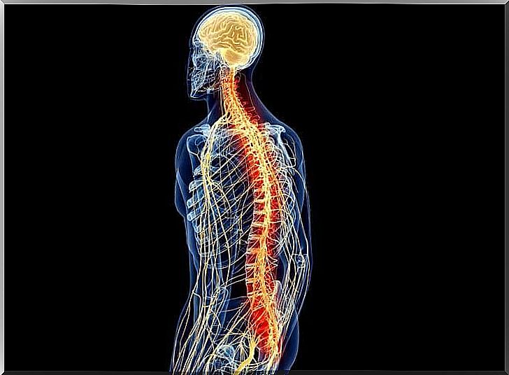
Spinal Cord: Anatomy And Physiology
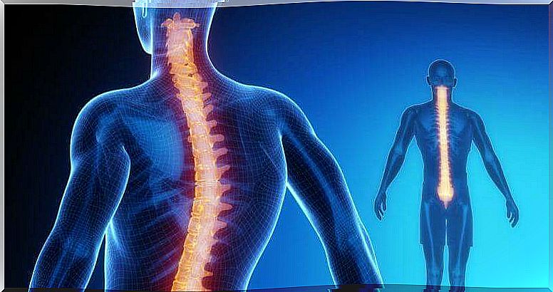
The spinal cord is part of the central nervous system (CNS) along with the brain. Its extension extends from the occipital foramen of the skull to approximately the first lumbar vertebra.
Along the spinal cord, 31 spinal nerves are connected. It is composed of a gray matter nucleus where neuronal bodies are located, which in turn is surrounded by a white matter where axons are located. Interestingly, the distribution of gray and white matter in the spinal cord is the opposite of that in the brain. On the other hand, as protection of the spinal cord there are the vertebrae, the supporting ligaments, the meninges and the cerebrospinal fluid.
The functions of the spinal cord are varied. On the one hand, it takes care of receiving and processing (in a superficial way) sensory information, and, on the other hand, of sending motor information from the brain. Its functions are essential and vitally important. An injury can cause serious effects such as motor paralysis or loss of sensation.
gray substance
Gray matter, unlike what occurs in the brain, is located in the inner part of the spinal cord. It is the place where neuronal bodies are located and where information is processed. It is configured in different horns: ventral, dorsal, lateral and intermediate.
- Dorsal horn: responsible for sensory information.
- Intermediate zone: it is the place where the interneurons that connect neurons to each other are found, they are association neurons.
- Lateral horn: found only at the thoracic and lumbar level. It is responsible for the body’s homeostasis, regulating the autonomic nervous system.
- Ventral horn: responsible for motor information.
Furthermore, within this gray matter there are several nuclei with different functions:
- I-IV: responsible for exteroceptive sensations. They register the sensations they receive from external stimuli, such as light, for example.
- V-VI: responsible for proprioceptive sensations. They report internally generated stimuli.
- VIII: performs the relay between the midbrain and the cerebellum. It is where neurons from the midbrain take over to the cerebellum and vice versa.
- IX: main motor area. It is where the descending neuronal bodies that come from the motor cortex conduct movement impulses.
- X: nucleus that surrounds the central duct and contains glial cells, which are support or support neurons.
The gray matter of the spinal cord is the place of replacement for motor and sensory information, but it also has to make a quick judgment about the information before it even reaches its destination. The latter is necessary to activate the reflex mechanism in emergency situations, such as when receiving a very painful stimulus.
white substance
The white matter of the spinal cord is where the fibers (axons) that send information both ascending and descending are located. Its main function is to send information. Like the substantia nigra, it is also divided into different parts, in this case columns:
- Back column : the one that sends somatic information.
- Ventral and lateral column : these are the efferent pathways that are responsible for sending information from the brain to the muscles. They are part of the motor system.
Within the white matter there are different pathways, ascending and descending. The tracts or fascicles bear the name of the two structures between which information circulates, and each of the areas sends different information.
- Gracile and cuneiform: responsible for touch and discriminative hand movements.
- Anterior and posterior spinocerebellar: unconscious movements from muscles, skin joints and subcutaneous tissue.
- Spino-olivary tract: although this tract has been located, its exact function is not known.
- Lateral spinothalamic: painful and thermal sensations.
- Spinotectal: afferent information for spinovisual reflexes.
- Spinothalamic anterior: light touch and pressure.
- Anterior and lateral corticospinal: gives agility and speed to movements.
- Spinal roof: participates in movements for visual stimuli.
- Vestibulospinal: responsible for maintaining balance.
- Olivo-spinal: influences the activity of motor neurons.
- Rubrospinal: inhibits the activity of the extensor muscles.
Thus, the white matter of the spinal cord is responsible for transmitting motor and sensory information in a wide range of movements and sensations, communicating multiple areas.
Ascending pathways (sensory)
The ascending pathways, as the name implies, are responsible for sending the information collected by the external senses (exteroceptive information) or internal stimuli (proprioceptive) to the cerebral cortex, where deeper processing will take place. Most ascending pathways are prominent in the thalamus, with the exception of olfactory stimuli that go straight to the olfactory bulb.

They are ascendant, centripetal, born from the periphery and provide information to the higher centers. Some of the nerve fibers serve to connect different segments of the spinal cord, while others ascend from the cord to higher centers and thus connect the spinal cord to the brain. These pathways carry information that may or may not reach consciousness.
In its simplest form, the pathway that ascends to consciousness consists of three neurons, but some afferent pathways use more or less. Many of the neurons present in the ascending pathways branch out and others participate in reflex muscle activity.
These are the pathways that carry information from the somatoreceptors. There are two main ways:
- Nocioceptive pathway, which conveys information about pain and temperature;
- Mechanical way, which transmits information about superficial and deep touch, proprioception and vibration.
Downhill (engine)
Pyramidal pathways are those descending (motor) nerve pathways that pass through the pyramids. They are responsible for fast, agile, fine and precise voluntary movement. There are three neurons involved in sending information to execute a movement. They follow the following circuit:
- Neuron 1: neuron located in the prefrontal and premotor cortex.
- Neuron 2: not always present in the pathway. It is an interneuron or internuncial neuron.
- Neuron 3: located in the anterior horn of the spinal cord. All pyramidal pathways end up showing contralaterality, which means that an injury to the right motor cortex will cause an injury to the left part of the body, for example.
The extrapyramidal pathway is responsible for involuntary movements, it comes from some subcortical structure that travels to the spinal cord. It regulates the execution of involuntary movements (walking, posture, muscle tone, alertness and instinctual behavior). Unlike the pyramidal system, it does not start in the cerebral cortex, but in various subcortical structures.
Another essential function of the descending motor pathways is to modulate reflex circuits in the spinal cord. The adaptability of spinal reflexes can change depending on the behavioral context, as sometimes the gain (strength) or even the sign (extension vs. flexion) of a reflex must be altered for the movement to adapt to the circumstances. The descending pathways are responsible for controlling these variables.
spinal cord reflexes
There are movements that we perform unconsciously, before the sensory information of the stimulus that causes the movement reaches the brain. These are reflex movements, such as removing your hand from a source of pain (called a lighter, for example), or closing your eyes when you hear a loud noise; we don’t control them.
The reflex is the simplest circuit in the nervous system. It starts at the receptors, which are structures that transform the energy of the stimulus into an electrical change in the peripheral afferent nerves that carry impulses to the integration center, the interneuron. The information passes to the efferent motor neurons so that the effector (muscle) performs reflex movement.
These movements occur thanks to the reflex arc. The neuron soma is located in the posterior root ganglion, passes through the dorsal, where it communicates with the interneuron, which is the association neuron that integrates the information and passes to the motor neuron in the ventral horn to exit through the ventral root and direct the nerve impulse to the muscle for its contraction.
Bibliography
- Carlson R. (2014). Physiology of the conduct . Madrid, Spain: Pearson education
- Tortora GJ, Derrickson B.(2013). Principles of anatomy and physiology . Buenos Aires: Panamerican Medical Editorial
- Kandell ER, Schwartz JH and Jessell TM (2001). Principles of Neuroscience . Madrid: McGraw-Hill/Interamericana.